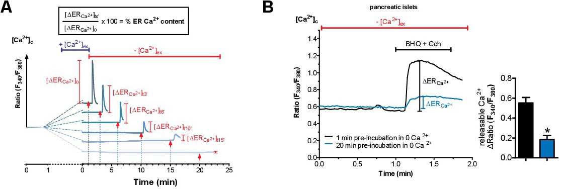
Fig. 1. Isolated pancreatic islets and β-cells have an atypical ER leak that is independent of SERCA activity. (A) Schematic representation of the ER Ca2+ leakage protocol (original traces were from a respective experiment with INS cells). After loading cells with the cytosolic Ca2+ indicator Fura-2/AM, they were perfused with Ca2+ containing EB for 1 min before switching to Ca2+-free buffer for predefined periods of time i.e. 1, 3, 6, 10, 15 and 20 min. For evaluation of ER Ca2+ content, ER Ca2+ stores were fully depleted by applying IP3-generating agonists (100 µM carbachol for pancreatic islets/β-cells or histamine for non-β-cells) together with 15 µM of the SERCA inhibitor BHQ - to avoid refilling of the ER - at the time points indicated with an arrow. The maximal ER store depletion was measured as maximal releasable ER Ca2+ in the cytosol whereas the ER is considered as fully filled at the one min time point and used as reference for calculating ER Ca2+ content. (B) Representative curves showing ER Ca2+ content indirectly measured with Fura-2/AM in isolated pancreatic islets. Islets were kept in Ca2+-free buffer for 1 min (black line) or 20 min (blue line) prior to ER Ca2+ store depletion by applying the SERCA inhibitor BHQ (15 µM) together with carbachol (Cch, 100 µM). Bars on the right represent corresponding statistics. *p<0.05 tested with unpaired Student's t-test, n≥4.